SAMACHEER CLASS 10 UNIT 13 NOTES
UNIT
13
SAMACHEER SCIENCE
CLASS 10
BIOLOGY
STRUCTURAL ORGANISATION OF ANIMALS
INTRODUCTION
·
Kingdom Animalia – 2 groups
·
Invertebrates
·
Vertebrates
·
Structural morphology & anatomy of an Invertebrate
(Leech) & a Vertebrate (Rabbit)
LEECH
·
Hirudinaria granulosa
·
Phylum – Annelida
·
Annelids – metamerically segmented worms
·
Well developed organ system
RABBIT
·
Oryctolagus cuniculus
·
Phylum – Chordata
·
Class – Mammalia
·
Mammals – highest group in animal kingdom – shows
advancements over other group
·
Warm blooded
·
Body covered by hair
·
Females – mammary gland – important feature
THE INDIAN CATTLE LEECH (Hirudinaria granulosa)
HABIT & HABITAT
·
Found in – India, Bangladesh, Pakistan, Myanmmar &
Srilanka
·
Lives in – Fresh water ponds, lakes, swamps & slow
streams
·
They are Ectoparasitic – lives on or in the
skin but not within the body
·
Feed on blood of fishes, frogs, cattle & human
·
Sanguivorous in nature – Blood sucking
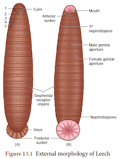
EXTERNAL MORPHOLOGY
SHAPE & SIZE
·
Body – soft, vermiform, elongated & segmented
·
Ribbon shaped – extended; Cylindrical – contracted
·
Grow to a length of 35cm
COLOURATION
·
Dorsal surface – Olive green
·
Ventral surface – Orange yellow
SEGMENTATION
·
Segmentation – metamerism (linear series of
body segment)
·
Body – metamerically divided into 33 segments
·
Segments – arranged one behind the other
·
Each segment – further superficially subdivided into
rings or annuli
·
Temporary Clitellum – segments 9 to 11 – to
produce cocoon during breeding season
RECEPTORS
·
Dorsal side – 5 pairs of eyes – on first 5 segments
·
Each segment – bears number of sensory projections –
receptors
·
Annular receptors – located in each annulus
·
Segmental receptors – located on the first annulus of
each segment
SUCKERS
·
Have 2 suckers
·
Sucker at anterior end – anterior sucker or oral
sucker – ventral in position – occupy 1st five segments
·
Posterior sucker – formed by – fusion of last 7
segments
·
Anterior sucker – helps in feeding
·
Both suckers – helps in attachment & locomotion
EXTERNAL APERTURES
·
MOUTH – located in the middle of anterior sucker
·
ANUS – a small aperture – opens on mid-dorsal of 26th
segment
·
NEPHRIDIOPORES – Nephridia – open to exterior – by 17
pairs of Nephridiopores – lie ventrically on the last annulus of each segment –
6 to 22
·
MALE GENITAL PORE – mid-ventral opening – between 2nd
& 3rd annuli of 10th segment
·
FEMALE GENITAL PORE – mid-ventrally – between 2nd
& 3rd annuli of 11th segment
DIVISIONS OF THE BODY
BODY WALL
·
Body wall – 5 layers
·
Cuticle – Outermost layer
·
Epidermis – lies below cuticle
·
Dermis – lies below epidermis – formed of connective
tissue
·
Muscular layer – formed of circular & longitudinal
muscles
·
Botryoidal tissue – lies below longitudinal muscles –
fills entire coelom around the gut
LOCOMOTION
·
Locomotion takes place by
·
Looping or crawling movement
·
Swimming movement
LOOPING/ CRAWLING MOVEMENT
·
By contraction & relaxation of muscles
·
2 suckers – for attachment – during movement on
substratum
SWIMMING MOVEMENT
·
Swim actively
·
Perform undulating movements
DIGESTIVE SYSTEM
·
Includes long alimentary canal & digestive glands
ALIMENTARY CANAL
·
Straight tube – mouth to anus
·
Mouth – triradiate aperture – middle of anterior
sucker – leads into small buccal cavity
·
Wall of buccal cavity – 3 jaws – single row of minute
teeth
·
Jaws – have papillae – bear openings of salivary
glands
·
Mouth & buccal cavity – occupy first 5 segments
·
Buccal cavity – leads into muscular pharynx
·
Surrounded by salivary glands
·
Saliva – contains hirudin – prevents coagulation of
blood
·
Pharynx – leads to crop – through short & narrow
oesophagus
·
Crop – largest portion of alimentary canal
·
Divided into – series of 10 chambers
·
Chambers – communicate with one another – through
circular apertures – surrounded by sphincters
·
Each chamber – pair of lateral, backwardly directed
caecae arises – as blind outgrowth – Caeca or Diverticula
·
Crop & diverticula – store large amount of blood –
which can be slowly digested
·
Last chamber of crop – opens into stomach
·
Stomach – leads into intestine – small straight tube –
opens into rectum
·
Rectum – opens to the exterior – by Anus
FOOD FEEDING & DIGESTION
·
Leech – feeds by sucking blood – cattle &n
domestic animals
·
While feeding – attaches to victim strongly – by
posterior sucker
·
Leech – triradiate or Y shaped incision – in skin of
host – by jaws – protruded through mouth
·
Blood sucked – by muscular pharynx – salivary
secretion poured in
·
Ingested blood – stored in crop & diverticulum
·
Blood passes – from crop into stomach
·
Digestion – takes place in stomach – by action of
proteolytic enzyme
·
Digested blood – absorbed slowly by the intestine
·
Undigested food – stored in rectum – egested through
anus
·
Leeches prevent blood clotting – secretes protein –
hirudin
·
They inject an anaesthetic substance – prevent host
from feeling their bite
DIGESTIVE SYSTEM
MORE TO KNOW
·
Leeches – no ear – sense vibrations through their skin
·
Have 2 to 10 tiny eyes – helps to locate food
·
Suck blood – 5 times more than their body weight
·
May take more than a year – for complete digestion
& absorption of a full meal
SEGMENTATION OF LEECH
RESPIRATORY SYSTEM
·
Respiration – through skin
·
Dense network of capillaries (tiny blood vessels) –
containing haemocoelic fluid – extends between epidermal cells
·
Exchange of gases – through diffusion
·
O2 dissolved in water – diffuses through
skin – into haemocoelic fluid – CO2 diffuses out
·
Skin – kept moist & slimy – by mucus secretion –
prevents from drying
CIRCULATORY SYSTEM
·
In Leech – circulation – through haemocoelic system
·
No true blood vessels
·
Blood vessels – replaced by channels – haemocoelic
channels or canals – filled with blood like fluid
·
Coelomic fluid – contains haemoglobin
·
There are 4 longitudinal channels
·
1 lies above alimentary canal (dorsal)
·
1 below alimentary canal (ventral)
·
Other 2 – lie on either side of alimentary canal
(lateral) – serve as heart & have inner valves
·
All 4 channels – connected together – posteriorly – in
26th segment
NERVOUS SYSTEM
·
CNS of Leech – consists of
·
A Nerve ring
·
a paired ventral nerve cord
·
Nerve ring – surrounds pharynx – formed of
·
Suprapharyngeal ganglion (Brain)
·
Circumpharyngeal connective &
·
Subpharyngeal ganglion
·
Subpharyngeal ganglion – lies below pharynx – formed
by – fusion of 4 pairs of ganglia
EXCRETORY SYSTEM
·
Excretion – through Nephridia – 17 pairs
·
Segmentally arranged – 6th to 22nd
segments
·
Nephridia – opens out by Nephridiopores
REPRODUCTIVE SYSTEM
·
Leech – Hermaphrodite – both male & female
reproductive organs – present in same animal
MALE REPRODUCTIVE SYSTEM
·
11 pairs of testes – arranged segmentally – 12 to 22
segments
·
Form spherical sacs – called Testes sacs
·
From Testes – arise a short duct – Vas efferens –
joins with Vas deferens
·
Vas deferens – becomes convoluted – to form Epididymis
or Sperm vesicle – stores spermatozoa
·
Epididymis – leads to – short duct – ejaculatory duct
·
Ejaculatory ducts on both sides – join to form –
genital atrium
·
Atrium – 2 regions
·
Coiled Prostate gland
·
Penial sac – consisting Penis – opens through the male
genital pore
FEMALE REPRODUCTIVE SYSTEM
·
Consists of – Ovaries, Oviducts & Vagina
·
Single pair of Ovary – 11th segment –
ventral side
·
Ovary – ribbon-shaped structure
·
Ova – budded off from ovary
·
From each ovary – a short oviduct arises
·
Oviducts – from both sides – joins together – form
common oviduct
·
Common Oviduct – opens into Vagina – pear-shaped –
lies mid-ventrally – in the posterior part of 11th segment
DEVELOPMENT
·
Internal Fertilization
·
Followed by – Cocoon formation
·
Cocoon / egg case – formed around 9th , 10th
& 11th segments
·
Development – direct
·
Takes place in cocoon
·
Cocoon – has 1 to 24 embryos
·
Young leech – resembling adults emerges
MORE TO KNOW – MEDICINAL VALUE OF LEECH
·
Leeches – effective – increasing blood circulation
& breaking blood clots
·
Used to treat Cardiovascular diseases
·
Leech saliva – biochemical substances – used for
preparation of drugs – treat hypertension (High BP)
PARASITIC ADAPTATIONS OF LEECH
·
Leech – parasitic mode of life – by sucking blood of
vertebrates – shows several important adaptations in their structure
·
Blood – sucked by pharynx
·
Anterior & posterior suckers – helps them to
attach to the host
·
3 jaws – inside mouth – causes painless Y shaped wound
– in the host’s skin
·
Salivary gland – hirudin – does not allow blood clot –
maintains continuous flow of blood
·
Parapodia & Setae – completely absent
·
Blood – stored in Crop – gives nourishment for several
months – therefore no secretion of digestive juices & enzymes
RABBIT (Oryctolagus cuniculus)
HABIT & HABITAT
·
Rabbits – gentle & timid animals
·
Leaping movements; Live in burrows
·
Distributed throughout the world
·
Herbivorous – feeds on – grass & vegetables –
turnips, carrots & lettuce
·
Rabbits – gregarious animals (moving in groups)
EXTERNAL MORPHOLOGY
SHAPE, SIZE & COLOURATION
·
Elongated & cylindrical body
·
Males & females – same size
·
Adults – 45 cm in length; 2.25 kg in weight
·
Colour – varies – white to black & white
·
Body – covered with fur – keeps it warm
BODY – DIVISION
·
Body – divisible into – Head, Neck, trunk & tail
HEAD
·
Head – ovoid, flattened & bears a truncate snout
·
Contains – mouth, external nares, eyes, ears &
vibrissae
·
Mouth – transverse slit-like – bounded by upper lip
& lower lip
·
Above mouth – 2 oblique openings – Nostrils
·
From each side of the upper lip – tactile hairs or
Vibrissae (Whiskers) – projects outwards
·
A pair of large, movable external ear / pinnae – on
top of the head
NECK
·
Connects head & trunk
·
Helps to turn the head
TRUNK
·
Divisible into
·
Anterior thorax
·
Posterior abdomen
·
Females – between ventral side of thorax & abdomen
– 4 or 5 teats or nipples are present
·
Bears – 2 pairs of pentadactyl limbs
·
Forelimbs – shorter than hindlimbs
· All the digits – have claws
·
Anus – present at – posterior end of the abdomen –
base of the tail
·
Females – ventral side – slit like – vulva present
·
Males – ventral side of anus – penis present
·
Males – pair of testes – enclosed by – Scrotal sacs
TAIL
·
Tail – short
·
Give signals to other rabbits – during danger
INTEGUMENT (SKIN)
·
Forms outer covering of the body
·
Structures derived from it – hairs, claws, nails &
glands (sweat, sebaceous & mammary glands)
·
Mammary glands – modified glands of the skin – secrete
milk – nourishes young ones
·
Sweat glands & sebaceous glands – embedded in the
skin – regulates body temperature
COELOM (BODY CAVITY)
·
Rabbit – Coelomate animal
·
Body – divided into – thoracic cavity & abdominal
cavity – separated by diaphragm (transverse partition)
·
Diaphragm – characteristic feature of mammals
·
Breathing movements – brought by – movement of
diaphragm
·
Lungs & heart – thoracic cavity
·
Digestive & Urinogenital system – abdominal cavity
· Includes – alimentary canal & associated digestive glands
·
Alimentary canal – consists of – Mouth, buccal cavity,
pharynx, oesophagus, stomach, small intestine, caecum, large intestine &
anus
·
Mouth – transverse slit – bounded by upper & lower
lips
·
Mouth – leads to buccal cavity
·
Floor of buccal cavity – muscular tongu
·
Jaws – bears teeth
·
Buccal cavity – leads to – Oesophagus – through
pharynx
·
Oesophagus – opens into stomach – followed by small
intestine
·
Caecum – thin walled sac – present at the junction of
small intestine & large intestine
·
Caecum – contains bacteria – helps in digestion of cellulose
·
Small intestine – opens into large intestine – has
colon & rectum
·
Rectum – opens outside – by anus
DIGESTIVE SYSTEM
DIGESTIVE GLANDS
·
Digestive glands – salivary glands, gastric glands,
liver, pancreas & intestinal glands
·
Digestive gland secretions – helps digestion of food –
in alimentary canal
DENTITION IN RABBIT
·
Teeth – hard bone-like structure – used to – cut, tear
& grind – food materials
·
2 sets of teeth – existence of 2 sets of teeth in the
life of an animal – called Diphyodont dentition
·
2 types of teeth – milk teeth (young ones) &
permanent teeth (Adults)
·
Rabbit – different types of teeth – Hence, dentition –
called heterodont
·
4 kinds of teeth in mammals – viz., Incisors (I),
Canines (C), Premolars (PM) & Molars (M)
·
Expressed – in Dental formula
·
Dental formula – simple method of representing the
teeth of a mammal
·
Number of each kind of tooth – in upper & lower
jaw – on one side is counted
·
Dental formula –
· Rabbit – Dental formula
·
Canines absent
·
Gap between Incisors & premolar – called diastema
·
Diastema – helps in mastication & chewing of food
– in herbivorous animals
RESPIRATORY SYSTEM
·
Respiration – takes place by – pair of lungs – light
spongy tissues – in thoracic cavity
·
Thoracic cavity – bound dorsally by – Vertebral
column; ventrally – sternum; laterally – ribs
·
Lower side of thoracic cavity – diaphragm – dome
shaped
·
Each lung – double membrane – pleura
·
Atmospheric air à External nostril à Nasal passage Pharynx
glottis wind pipe
·
Anterior part of wind pipe – enlarged – form larynx /
voice box – its wall supported by 4 cartilagenous plates
·
Inside larynx – vocal cord – its vibrations – produces
sound
·
Larynx – leads to Trachea / Wind pipe
·
Tracheal walls – supported by – rings of cartilage –
helps in free passage of air
·
Epiglottis – prevents entry of food – into trachea
through glottis
·
Trachea – divides into 2 branches – Bronchi – enters
lungs – divides into further branches – Bronchioles – end in alveoli
·
Respiratory events – Inspiration (breathing in) &
Expiration (breathing out) – allows exchange of gases (O2 & CO2)
·
Inspiration – active process; Expiration – passive
process
RESPIRATORY SYSTEM
CIRCULATORY SYSTEM
·
Circulatory system – formed by – blood, blood vessels
& heart
·
Heart – pear shaped – lies in thoracic cavity –
between lungs
·
Heart – enclosed by Pericardium (double layered membrane)
·
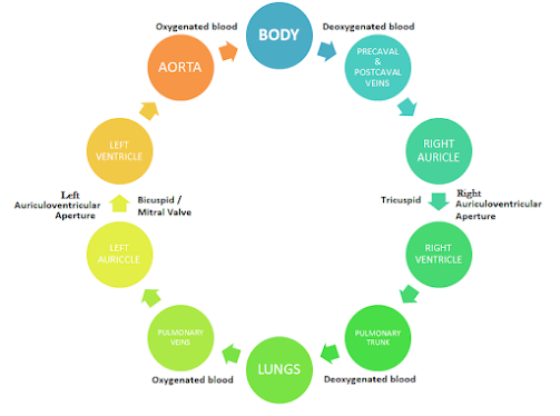
Heart – 4 chambers – 2 auricles; 2 ventricles
·
Right & left auricles – separated by –
Interauricular septum
·
Right & left ventricles – separated by –
Interventricular septum
·
Right auricle à opens into – right ventricle
– by right auriculoventricular aperture – guarded by tricuspid valve
·
Left auricle à opens into – left ventricle – by left auriculoventricular aperture –
guarded by bicuspid valve / mitral valve
·
Opening of pulmonary artery & aorta – guarded by 3
semilunar valves
·
Right auricle – receives deoxygenated blood – through
2 precaval (superior Vena cava) & post caval (inferior venacava) veins –
from all parts of the body
·
Left auricle – receives oxygenated blood – from
pulmonary veins – from lungs
·
From right ventricle – pulmonary trunk arises –
carries deoxygenated blood – to lungs
·
From Left ventricle – Aorta (systematic arch) arises –
supplies oxygenated blood – to all parts of the body
CIRCULATORY SYSTEM
NERVOUS SYSTEM
·
Nervous system – formed of
·
CNS – Central Nervous System
·
PNS – Peripheral Nervous System
·
ANS – Autonomic Nervous System
·
CNS – Brain & Spinal cord
·
PNS – 12 pairs of cranial nerves & 37 pairs of
spinal nerves
·
ANS – Sympathetic & Parasympathetic nerves
·
Brain – in cranial cavity – covered by 3 membranes
·
Outer Duramater
·
Inner Piamater &
·
Middle Arachnoid membrane
·
Brain – divided into
·
Forebrain (Prosencephalon)
·
Midbrain (Mesencephalon) &
·
Hindbrain (Rhombencephalon)
·
Forebrain – pair of olfactory lobes, cerebral
hemispheres & diencephalon
·
Right & left Cerebral hemisphere – connected –
Corpus callosum (transverse band of nerve tissue)
·
Mid brain – optic lobes
·
Hind brain – Cerebellum, Pons Varolii & Medulla
Oblongata
URINOGENITAL SYSTEM
·
Comprises – Urinary / Excretory system & genital /
reproductive system – hence described as Urinogenital system – in Vertebrates
EXCRETORY SYSTEM
·
Kidney – made of several Nephrons
·
Nephrons – separates nitrogenous wastes – from blood –
excretes as urea
·
Kidneys – dark red, bean shaped – in abdominal cavity
·
From each kidney – arises ureters – opens posteriorly
into urinary bladder – leads into urethra (muscular duct)
REPRODUCTIVE SYSTEM
·
Sexual dimorphism – seen in Rabbits – male &
female sexes – separate & morphologically different
MALE REPRODUCTIVE SYSTEM
·
Male Reproductive system – consists of – pair of
testes – ovoid shape
·
Testes – enclosed by scrotal sacs – in abdominal
cavity
·
Testes – has many fine tubules – seminiferous tubules
– this tubule network – lead into – epididymis (coiled tubule) – lead into Vas
deferens (Sperm duct) – joins in Urethra (below urinary bladder) – runs
backwards – passes into Penis
·
3 accessory glands – prostrate gland, cowper’s gland
& perineal gland – secretions involved in – Reproduction
FEMALE REPRODUCTIVE SYSTEM
·
Consists of – pair of ovaries – small ovoid – behind
the kidneys – in abdominal cavity
·
Oviduct arises from each ovary – through a funnel
shaped opening
·
Anterior part of oviduct – fallopian tube – leads to
wider tube – called Uterus – joins together – form median tube – Vagina
·
Common tube formed – union of urinary bladder &
vagina – urinogenital canal or vestibule – runs backwards – opens out by slit
like aperture – Vulva
·
Accessory glands – pair of cowper’s gland &
perineal gland – present


Comments
Post a Comment