SAMACHEER CLASS 10 UNIT 14 NOTES
UNIT
14
samacheer
SCIENCE
CLASS 10
BIOLOGY
TRANSPORTATION IN PLANTS & CIRCULATION IN ANIMALS
INTRODUCTION
·
Multicellular organisms – possess millions of cells
·
Every cell – needs essential substances – nutrients
& oxygen – to maintain life & survival
·
Food – only source of energy
·
Each cell – get energy by breaking of glucose
·
Cells utilise this energy - & carries out various
activities
TRANSLOCATION
·
Water & mineral salts – absorbed by roots – reach
all parts of the plant – through Xylem
·
Food – synthesized by the leaves – translocated to all
parts of the plant – through Phloem
·
Bulk movement of substances – through vascular tissue
– called Translocation
·
Transport – means to carry things from one place to
another
·
In larger animals – transport of nutrients, salts,
oxygen, hormones & waste products – around the body – done by ‘Circulatory
system’
·
Circulatory system – consists of circulating fluids
(blood, lymph) & the heart & blood vessels – form the collecting &
transporting system
MEANS OF TRANSPORT
·
Transport of materials – in & out of cells –
carried out by – diffusion & active transport in plants
DIFFUSION
·
Movement of molecules – in liquid & solid – from
region of higher concentration – to region of lower concentration – without
using energy – Diffusion
·
This is a passive process

ACTIVE TRANSPORT
·
Uses energy to pump molecules – against concentration
gradient
·
Carried out by membrane bound proteins
·
These proteins – use energy to carry substances across
the cell membranes – called pumps
·
Pumps – transport substances – from low concentration
to high concentration – uphill transport
OSMOSIS
·
Movement of solvent or water molecules – from region
of higher concentration – to region of lower concentration – through
semi-permeable membrane
·
This process – carried out till – an equilibrium is
reached
·
Osmosis – passive movement of water / solvent
molecules

PLASMOLYSIS
· Occurs when water moves out of the cell – results in shrinkage of – cell membrane – away from cell wall

DEMONSTRATION OF OSMOSIS
·
A thistle funnel – mouth covered with semi-permeable
membrane
·
Funnel – filled with sucrose solution
·
Funnel – kept inverted in a beaker – containing water
·
Water – diffuse into funnel – across the membrane –
due to osmosis
·
Result – raise in the level of sucrose solution in the
funnel

IMBIBITION
·
A type of diffusion – in which a solid absorbs water –
swells up
·
Example: Absorption of water – by seeds & dry
grapes
·
If there is no imbibition – seedlings would not emerge
out of the soil

ROOT HAIR
·
Millions of root hairs – tips of roots – absorb water
& minerals – by diffusion
·
Root hairs – thin walled, slender – extension of
epidermal cells – that increase surface area for absorption

PATHWAY OF WATER ABSORBED BY ROOTS
·
Once water enters root hairs – concentration of water
becomes more – than its cortex
·
So, water from root hairs – moves to cortex (cortical
cells) – by osmosis
·
Water – then reaches xylem
·
Xylem – transports water to stem & leaves

HOW DO PLANTS ABSORB WATER?
·
Water – absorbed along with minerals – by root hairs –
through diffusion
·
Once water is absorbed by root hairs – it moves deeper
into root layers – by 2 pathways
·
(i) Apoplast Pathway
·
(ii) Symplast Pathway
APOPLAST PATHWAY
·
Apoplastic movement of water – occurs through –
intercellular spaces & the cell walls
·
Does not involve – crossing the cell membrane
·
Movement – dependent on the gradient – (Higher
concentration to lower concentration)

SYMPLAST PATHWAY
·
Symplastic movement – water travels through cells –
i.e., through cytoplasm
·
Intercellular movement – through plasmodesmata
·
As water moves through cytoplasm – movement is slower
·
Movement – down a potential gradient (Water potential
– tendency of water to move from one place to another – due to osmosis,
gravity, etc.,)
TRANSPIRATION
·
Transpiration – evaporation of water in plants –
through stomata (leaves)
·
Stomata – open (Day); closed (night)
·
Opening & closing of stomata – due to turgidity of
guard cells
·
Turgidity (increases) – stoma opens
·
When guard cells lose water (turgidity decrease) –
stoma closes

TRANSPIRATION PULL
·
Water evaporates from – Mesophyll cells – through
stomata
·
Water concentration – lowers in mesophyll cells
·
Result – water from xylem (veins) à drawn into mesophyll cells (through osmosis)
·
As water is lost from leaves – pressure created at top
– this pulls more water from xylem to mesophyll cells – process called
‘Transpiration Pull’
·
This pressure – extends upto roots – cause the roots
to absorb more water from soil – ensures continuous flow of water – from roots
to leaves
FACTORS THAT AFFECTS TRANSPIRATION
EXTERNAL FACTORS
·
Temperature
·
Light
·
Humidity
·
Wind speed
INTERNAL FACTORS
·
Number of stomata
·
Percentage of open stomata
·
Water status of plant
·
Canopy structure (organisation/spatial arrangement of
a plant)
IMPORTANCE OF TRANSPIRATION
·
Creates transpirational pull for – transport of water
·
Supplies water for photosynthesis
·
Transports minerals – from soil to all plant parts
·
Cools leaf surface – by evaporation
·
Keeps cells turgid – maintains their shape
ROOT PRESSURE
·
As ions from soil – actively transported into xylem of
the root – water moves and creates pressure inside xylem – called root pressure
·
Root pressure – responsible for pushing water – to
smaller height in the stem
UPTAKE OF MINERALS
·
Plants – depend on minerals in the soil – for its
nutrition
·
All minerals – cannot be passively absorbed by roots
·
2 factors for this
·
(i) Minerals in soil – charged particles (ions) –
cannot move across cell membrane
·
(ii) Concentration of minerals in soil – lower than
their concentration in the root
·
Therefore, most minerals enters root by – active
absorption – through cytoplasm of epidermal cells
·
This process – needs energy (ATP)
·
Absorbed minerals – transported to all parts – by
transpiration pull
TRANSLOCATION OF MINERAL IONS
·
Minerals – remobilised from older dying leaves – to
younger leaves
·
This phenomenon – seen in – Deciduous plants
·
Elements – P, S, N & K – easily remobilised
·
Elements – Ca – not remobilised
·
Small amount of material – exchange between xylem
& phloem
PHLOEM TRANSPORT
·
Food – produced in leaves – transported by phloem – to
area of requirement / stored
·
Phloem tissue – composed of sieve tubes – have sieve
plates – cytoplasmic strands pass through the pores in sieve plates
·
Phloem – transport food (sucrose) – from source to
sink
·
Source – where food is synthesized (leaves)
·
Sink – where is needed / stored
·
Source & sink – may be reversed – depending on
season / plant’s need
·
Since, source-sink relationship – variable – direction
of movement in phloem – can be upwards / downwards – Bidirectional
·
Movement in xylem – always upwards – Unidirectional
TRANSLOCATION OF SUGARS
·
Translocation of sugars – from source to sink –
through pressure flow hypothesis
·
Glucose at source (prepared by photosynthesis) –
converted to sucrose
·
Sucrose – moves to companion cells – then into living
phloem sieve tube cells – by active transport
·
This process creates – hypertonic condition in phloem
(water content less)
·
Water from adjacent xylem – moves into phloem – by
osmosis
·
Osmotic pressure builds up – makes phloem sap moves to
– area of lower pressure
·
By active transport – sucrose moves to places – where
its needed / stored
·
As sugars moved – osmotic pressure decreases – water
moves out of phloem
ASCENT OF SAP & ITS EVENTS – AN OVERVIEW
·
Upward movement of water & minerals – from roots
to different plant parts – called ascent of sap
·
Many factors play a role in this

STEPS INVOLVED
·
ROOT PRESSURE
·
Water from soil – enters root hairs – by osmosis
·
Root pressure – moves water upwards – up to base of
stem
·
CAPILLARY ACTION
·
Water / any liquid – rise in capillary tube – due to
physical force – phenomenon called Capillary action
·
Water in stem – rises up to certain height – due to
capillary action
·
ADHESION-COHESION OF WATER MOLECULES
·
Water flows continuously in xylem – due to adhesion
& cohesion forces among molecules
·
COHESION
·
force of attraction between water molecules
·
ADHESION
·
force of attraction between molecules of different
substances
·
Water molecules – stick to xylem – due to adhesion
force
·
TRANSPIRATION PULL
·
Transpiration through stomata – creates vacuum –
creates suction – called Transpiration pull
·
Transpiration pull – sucks water in xylem tubes –
makes water to rise to great heights – even in tall plants
MORE TO KNOW
·
Dews – droplets of water – on leaves of grass – seen
early morning – climate is humid & excess water in soil
·
This is due to root pressure – phenomenon called
Guttation
·
Takes place through – special cells – Hydathodes

DEMOSTRATION OF ROOT PRESSURE
·
Choose – small soft stemed plant
·
Cut the stem – horizontally near the base – in the
morning
·
Drops of solution – oozes out of the cut stem
·
This is due to root pressure
BLOOD
·
Blood – main circulatory medium – in our body
·
Red coloured fluid – connective tissue
COMPONENTS OF BLOOD
·
Blood – 2 main components
·
(i) Fluid plasma
·
(ii) Formed elements (blood cells) – suspended in
plasma
(i) PLASMA
·
Slightly alkaline, non-living, intercellular substance
·
Constitute 55% of blood
·
Organic substances – proteins, glucose, urea, enzymes,
hormones, vitamins & minerals – present in plasma
(ii) FORMED ELEMENTS OF BLOOD
·
Blood corpuscles – 3 types
·
(1) Red Blood Corpuscles (RBC) or Erythrocytes
·
(2) White Blood Corpuscles (WBC) or Leucocytes
·
(3) Blood Platelets or Thrombocytes
(1) RED BLOOD CORPUSCLES (ERYTHROCYTES)
·
Most abundant cells – in human blood
·
Formed in – Bone marrow
·
RBC’s – red colour – due to presence of respiratory
pigment – Haemoglobin
·
Matured Mammalian RBC’s – lack cell organelles &
nucleus
·
Biconcave & disc shaped
·
Life span – 120 days
·
RBC’s – transport O2 from lungs to tissues

WHY MAMMALIAN RBC LACK CELL ORGANELLES & NUCLEUS?
·
Lack nucleus – makes the cell biconcave – increase
surface area – for O2 binding
·
Loss of Mitochondria – allows RBC to transport all O2
to tissues
·
Loss of ER – makes RBC more flexible – to move through
narrow capillaries
(2) WHITE BLOOD CORPUSCLES (LEUCOCYTES)
·
WBC – colourless; Do not have Haemoglobin &
nucleated cells
·
Found in – bone marrow, spleen, thymus & lymph
nodes
·
Capable of amoeboid movement
·
WBC – grouped into 2 categories
·
(A) Granulocytes
·
(B) Agranulocytes
(A) GRANULOCYTES
·
Contain granules – in cytoplasm
·
Nucleus – irregular / lobed
·
3 types
·
(i) Neutrophils
·
(ii) Eosinophils
·
(iii) Basophils

(i) NEUTROPHILS
·
Large in size
·
Have 2 to 7 lobed nucleus
·
These corpuscles form 60 to 65% of total leucocytes
·
Number increases – during infection & inflammation
(ii) EOSINOPHILS
·
Bilobed nucleus
·
Constitute 2 to 3% of total leucocytes
·
Number increases – during allergy & parasitic
infections
·
Brings about – detoxification of toxins
(iii) BASOPHILS
·
Lobed nucleus
·
Form 0.5 to 1.0 % of total leucocytes
·
They release chemicals – during inflammation

(B) AGRANULOCYTES
·
Granules – not found in cytoplasm
·
2 types
·
(i) Lymphocytes
·
(ii) Monocytes
(i) LYMPHOCYTES
·
Forms 20 to 25% of total leucocytes
·
They produce antibodies – during bacterial & viral
infections
(ii) MONOCYTES
·
Largest of leucocytes
·
Amoeboid in shape
·
Form 5 to 6% of total leucocytes
·
They are phagocytic & engulf bacteria (Cells uses
plasma membrane to engulf large particles)
(3) BLOOD PLATELETS OR THROMBOCYTES
·
Small & colourless
·
Do not have nucleus
·
There are 2,50,000 – 4,00,000 platelets / cubic mm of
blood
·
Life span – 2 to 3 days
·
Helps in clotting of blood
·
Platelets – form clot at site of injury – prevents
blood loss
 |
| Platelet |
MORE TO KNOW
·
ANEMIA – Decrease in number of erythrocytes
·
LEUCOCYTOSIS – Increase in no. of Leucocytes
·
LEUKOPENIA - Decrease in no. of Leucocytes
· THROMBOCYTOPENIA - Decrease in no. of Thrombocytes
FUNCTIONS OF BLOOD
·
Transport of respiratory gases (O2 & CO2)
·
Transport of digested food to different body cells
·
Transport of hormones
·
Transport of nitrogenous wastes – Ammonia, urea &
uric acid
·
Involved in protection of body & defence against
diseases
·
Acts as buffer & helps in regulation of pH &
body temperature
·
Maintains proper water balance in the body
BLOOD VESSELS – ARTERIES & VEINS
·
Blood vessels – network of branched tubes – transports
blood
·
3 types of blood vessels – Arteries, Veins &
Capillaries
ARTERIES
·
Thick & elastic vessels
·
Carry blood away from heart to different body parts
·
All arteries carry oxygenated blood, except Pulmonary
artery (carry de-oxygenated blood to lungs)

VEINS
·
Thin & non-elastic vessels
·
Carry blood to the heart from different body parts
·
All veins carry de-oxygenated blood, except Pulmonary
veins (carry oxygenated blood from lungs to heart)
CAPILLARIES
·
Narrow tubes – formed by branching of arterioles –
unite to form venules & veins
·
8 µm – diameter
·
Formed of single layer of endothelial cells
DIFFERENCES BETWEEN ARTERY & VEIN

TYPES OF CIRCULATORY SYSTEM
·
Animals possess – 2 types of Circulatory system
·
(i) Open type
·
(ii) Closed type
(i) OPEN TYPE
·
Blood is pumped by heart – into blood vessels
·
Blood vessels – open into blood spaces – called
Sinuses
·
Sinuses are body cavities – called Haemocoel
·
Capillary system – absent
·
Example: Arthropods, Molluscs & Ascidians

(ii) CLOSED TYPE
·
Blood flows in a complete circuit – around the body –
through blood vessels
·
Blood flows – from arteries to veins – through
capillaries
·
Example: Vertebrates
MORE TO KNOW
·
Closed
circulatory system – discovered by – William Henry (1828) – Father of
Physiology
STRUCTURE OF HUMAN HEART
·
Heart – muscular pumping organ – pumps blood into
blood vessels
·
Human heart – situated between lungs – slightly tilted
towards left – above the diaphragm – in thoracic cavity
·
Heart – made of specialized muscle – Cardiac muscle
·
Heart – enclosed in – double walled sac – Pericardium
– contains lubricating fluid – Pericardial fluid
· Pericardial fluid – reduces friction during heart beat – protects from mechanical injury

CHAMBERS OF THE HEART
·
Human heart – 4 chambers
·
2 upper, thin walled chambers – auricle or atria
(sing. Atrium)
·
2 lower, thick walled chambers – Ventricles
·
Chambers separated by – partition – Septum
·
Septum between auricle & ventricle – prevents
mixing of oxygenated & deoxygenated blood

AURICLES
·
2 auricles separated by – interatrial septum
·
Left atrium – smaller than right
·
Right atrium – receives deoxygenated blood – from
different body parts – through main veins (Superior vena cava, Inferior vena
cava & coronary sinus)
·
Pulmonary Veins – brings oxygenated blood – to left
atrium – from lungs
·
Right & left auricles – pump blood to – right
& left ventricles respectively
VENTRICLES
·
Ventricles – lower part of the heart
·
2 ventricles – separated by – inter ventricular septum
·
Left & right ventricles – thick walled – because
they have to pump out blood with force away from heart
·
Right ventricle – arises – Pulmonary trunk –
bifurcates to form – right & left Pulmonary arteries
·
Right & left pulmonary arteries – supply
deoxygenated blood – to lungs of respective sides
·
Left ventricle – longer & narrower than right
ventricle
·
Left ventricle – walls 3 times thicker than right
ventricle
·
Left ventricle – arises – Aorta – supplies oxygenated
blood to different parts of the body
·
Coronary arteries – supply blood to heart
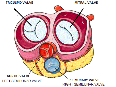
VALVES
·
Valves – muscular flaps – regulate flow of blood – in
single direction
·
Prevents back flow of blood
·
3 types of valves
·
(i) Right atrioventricular valve
·
(ii) Left atrioventricular valve
·
(iii) Semilunar valves

(i) RIGHT ATRIOVENTRICULAR VALVE
·
Located between – right auricle & right ventricle
·
Has 3 thin triangular leaf like flaps – called
Tricuspid valve
·
Apices of flaps – held in position by – Chordae
Tendinae – arise from – muscular projection in ventricle wall – called
Papillary muscles


(ii) LEFT ATRIOVENTRICULAR VALVE
·
Located between – Left auricle & left ventricle
·
Has 2 cusps – called Bicuspid / Mitral valve
(iii) SEMILUNAR VALVE
·
Major arteries (Pulmonary artery & Aorta) – which
leave the heart – have semilunar valves
·
They present the back flow of blood into ventricles
·
They are Pulmonary & Aortic semilunar valves
MORE TO KNOW
·
Heart chambers in Vertebrates
·
Two chambered – Fishes
·
Three chambered – Amphibians
·
Incomplete four chambered – Reptiles
· Four chambered – Aves, Mammals & crocodiles (reptiles)
TYPES OF BLOOD CIRCULATION
·
Blood circulates – as oxygenated & deoxygenated
blood
·
Types of circulation
·
(i) Systematic Circulation
·
(ii) Pulmonary Circulation
·
(iii) Coronary Circulation
(i) SYSTEMATIC CIRCULATION
·
Circulation of oxygenated blood – from left ventricle
– to various body organs – through Aorta
·
Return of deoxygenated blood – to right Atrium
(ii) PULMONARY CIRCULATION
·
Circulation starts in – right ventricle – deoxygenated
blood is carried by – pulmonary artery – to lungs
·
Pulmonary veins – collect oxygenated blood from lungs
– supplies to left atrium

(iii) CORONARY CIRCULATION
·
Supply of blood – to heart muscles (Cardiac muscles)-
Coronary circulation
·
Cardiac muscles – gets oxygenated blood from –
coronary arteries – originate from Aortic arch
· Deoxygenated blood from Cardiac muscles – drains into right atrium – by coronary sinuses

CIRCULATION
DOUBLE CIRCULATION
·
When blood circulates through the heart – one complete
cycle – called Double Circulation
·
Here oxygenated blood & deoxygenated blood – do
not mix
SINGLE CIRCULATION
·
In some animals – oxygenated & deoxygenated blood
mix – pass through heart only once – called Single Circulation
HEART BEAT
·
One complete contraction (systole) & relaxation
(diastole) – of atrium & ventricle of heart – Heart beat
·
Normal heart beat – 72 to 75 times per minute
MORE TO KNOW
Neurogenic Heart Beat
·
Initiated by – nerve impulse – from a nerve ganglion –
near the heart
·
Example: Annelids, most Arthropods
Myogenic Heart Beat
·
Initiated by – specialized group of modified heart
muscles
·
Example: Mollusca, Vertebrates
CONDUCTION OF HEART BEAT
·
Human heart – myogenic in nature
·
Contraction – initiated by – Sino-atrial (SA) node
(special portion of heart muscle)
·
SA node – present in the wall of right atrium – near
opening of Superior vena cava
·
SA node – broader at top; tapering below – made of
thin fibres
·
SA node – acts as ‘Pacemaker’ of heart – as it is
capable of initiating impulse – stimulates heart muscles to contract
·
Impulse from SA node – spreads as wave of contraction
– over right & left atrial wall – pushing blood through Atrioventricular
valves – into ventricles
· Wave of contraction from SA node – reach Atrioventricular (AV) node – stimulated to emit an impulse of contraction – spreads to ventricular muscle – via atrioventricular bundle & Purkinje fibers

DO YOU KNOW?
·
Atrioventricular bundle – discovered by HIS (1893) –
called Bundle of HIS
PULSE
·
When Heart beats – blood forced into arteries
·
Expansion of artery – every time when blood is forced
into – called Pulse
·
Felt by placing fingertip – on artery – near the wrist
·
Normal pulse rate – 70 to 90 /min
CARDIAC CYCLE
·
Sequence of events – from beginning to completion of
one heart beat – Cardiac cycle
·
During Cardiac cycle – blood flows through heart
chambers – in a specific direction
·
One cardiac cycle – lasts 0.8 second
·
Events during cardiac cycle involves
·
(a) Atrial Systole – contraction of auricles (0.1 sec)
·
(b) Ventricular Systole – contraction of ventricles
(0.3 sec)
· (c) Ventricular diastole – relaxation of ventricles (0.4 sec)

HEART SOUND
·
Rhythmic closure & opening of valves – causes
sound of the heart
·
First sound – LUBB – longer duration – produced by
closure of Tricuspid & Bicuspid valves – Ventricular Systole
·
Second sound – DUPP – shorter duration – produced by
closure of semilunar valves – at the end of Ventricular Systole
BLOOD PRESSURE
·
Force exerted – during blood flow – against the
lateral walls of arteries – Blood Pressure
·
BP – high in arteries & capillaries; Low in veins
·
BP – expressed in terms of – Systolic pressure &
diastolic pressure
SYSTOLIC PRESSURE
·
During ventricular systole – left ventricle contracts
– forces blood into Aorta
·
Pressure – rises to peak – Systolic Pressure
DIASTOLIC PRESSURE
·
During Diastole – ventricles relax – pressure falls to
the lowest value – Diastolic Pressure
BLOOD PRESSURE
·
Healthy adult – Normal systolic & diastolic
pressure – 120 mm/80mm Hg
·
BP varies – during physical exercise, anxiety,
emotions, stress & sleep
·
Prolonged or constant elevation of BP – Hypertension
(High BP) – increase the risk of heart attack & stroke
·
Decrease in BP – Hypotension (Low BP)
STETHOSCOPE
·
Used to detect – sounds produced by internal organs of
our body
·
Heart sound – heard by placing stethoscope – on chest
·
Useful diagnostic tool – to identify & locate
health problems & diagnose disease
· Modern Electronic Stethoscope – high precisioned instrument

SPHYGMOMANOMETER
·
Clinical instrument – used to measure BP – when the
person is in – relaxed & resting condition
·
Pressure of brachial artery – measured
·
Helps to diagnose – increased/decreased BP
· Monometric & modern digital types – used to measure BP

BLOOD GROUPS
·
Blood grouping concept – developed by – Karl
Landsteiner (1900)
·
He identified blood groups – A, B & O
·
AB blood group – recognised by – Decastello &
Steini (1902)
·
Human blood –
contains Agglutinogens or Antigens (Ag) & Agglutinins or Antibodies (Ab)
·
Ag – found on the membrane surface of RBC
·
Ab – present in plasma
BLOOD GROUPS
· Based on the presence of antigen & antibodies – human blood group is classified as – A, B, AB & O
·
An individual has one of the 4 blood groups
·
(i) ‘A’ GROUP individuals
·
Antigen A – present on RBC’s surface
·
Antibody b (anti-b) – present in plasma
·
(ii) ‘B’ GROUP individuals
·
Antigen B – present on RBC’s surface
·
Antibody a (anti-a) – present in plasma
·
(iii) ‘AB’GROUP individuals
·
Antigen A & B – present on RBC’s surface
·
Antibody – absent in plasma
·
(iv) ‘O’ GROUP individuals
·
Antigen A or B –
absent on RBC’s surface
·
Antibodies a & b (anti a & b)– present in
plasma

BLOOD DONATION
·
In blood transfusion – we must consider – antigen
& antibody compatibility (matching) – between donor & recipient
·
When the blood group is mismatched – it leads to
agglutination (clumping) – causes death
·
Persons with ‘AB’ blood group – Universal recipient –
can receive blood from any blood group
·
Persons with ‘O’ group – Universal Donor – can donate
blood to any blood group
RH FACTOR
·
Rh Factor – discovered by Landsteiner & Weiner
(1940) – in Rhesus Monkey
·
Antigen Rh – found on the surface of RBC’s
·
Rh+ (positive) persons – have Rh antigen on
the RBC’s surface
·
Rh- (negative) persons – donot have Rh
antigen on the RBC’s surface
·
Antibodies developed against Rh antigen – Rh
antibodies
LYMPHATIC SYSTEM
·
Lymphatic system consists of – lymphatic capillaries,
lymphatic vessels, lymph nodes & lymphatic ducts
·
Lymph – fluid that flows through lymphatic system
·
Lymphatic capillaries – unite to form large lymphatic
vessels
· Lymph nodes – small oval or pear shaped structures – located along lymphatic vessels

LYMPH
·
Lymph – colourless fluid – formed when plasma,
proteins & blood cells – escape into intercellular spaces – through pores
present in walls of capillaries
·
Lymph – from intercellular space – drains into
lymphatic capillaries
·
Similar to blood plasma – but colourless &
contains less proteins
·
Lymph – contains very less – nutrients, oxygen, CO2,
water & WBC
FUNCTIONS OF LYMPH
·
Supplies nutrition & O2 - to parts –
where blood cannot reach
·
Drains away – excess tissue fluid & metabolites
·
Returns – proteins to the blood from tissue spaces
·
Carries absorbed fats from small intestine to blood
·
Lymphatic capillaries of intestinal villi (Lacteals) –
absorb digested fats
·
Lymphocytes in Lymph – defend body from infections
Comments
Post a Comment